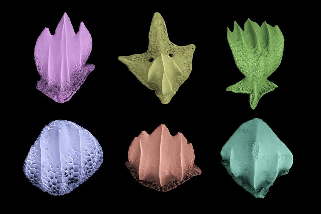
After visiting the Smithsonian’s National Museum of Natural History in May to excise small pieces of skin from across the bodies of nearly 40 specimens, representing 36 species and 16 families of sharks (nearly half of all the extant families), and meticulously treating the skin with Clorox to separate the individual denticles from the underlying tissue, here is a sneak preview of the final product under a scanning electron microscope. The denticle crown definition is quite stunning, right?

Scanning electron microscope (SEM) images of shark dermal denticles from the three main shark families represented in our sediment samples. A blacknose shark (Carcharhinus acronotus) denticle is shown for family Carcharhinidae, a scalloped hammerhead (Sphyrna lewini) denticle for family Sphyrnidae, and a nurse shark (Ginglymostoma cirratum) denticle for family Ginglymostomatidae. All of the denticles were isolated from pieces of skin excised from preserved museum specimens. Scale bar = 100µm.
And for the viewing pleasure of those more artistically-minded folks, a colorful rendition edited by my research advisor is shown below. Enjoy!
